Page 57 - MaquetaFundCIEN-2019-ENG
P. 57
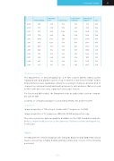
  3. Scientific activity
57
  FUNCTIONAL
  VOLUME- TRY
 ESPECTRA
DIFFUSION TENSOR
 MISCELLA- NEOUS
  ASL
  2008
  255
    339
   156
  178
   312
    259
  2009
337
  638
 866
423
 845
  326
  2010
  910
    852
   1042
  556
   1506
    305
  2011
1124
  845
 786
528
 1904
  398
  2012
  1858
    1512
   583
  1330
   2938
    949
  2013
 1436
   1276
  516
 1116
  2544
   950
  2014
880
  937
 367
847
 1920
  729
  2015
  828
    912
   309
  783
   1985
    669
  2016
739
  749
 341
618
 1661
  572
  2017
  614
    648
   299
  594
   1415
    523
  2018
487
  519
 226
124
 1051
  367
  2019
 473
   513
  306
 285
  1033
   278
   Provision of services
The Department of Neuroimaging has a 3T MR scanner (GEHC, HDxt) system equipped with dual gradient system of up to 50mt/m, 3 antennas for brain studies (transmitter/receiver quadrature antenna, receiving 8 channels antenna and 16 channels receiving antenna) and small antennas for rats and mice. Data is stored in PACS with direct recovery capacity for five years of work.
For Functional MRI studies, the Department has an audio/video system compati- ble with 3T MRI.
A variety of software packages is used, mainly SPM12, FSL and Freesurfer.
Sequences
Image acquisition of 3D isotropic studies with T1 sequences for VBM.
Image acquisition of T2 sequences, DWI, ASL, BOLD and spectroscopy.
The service provision data are publicly available on the CIEN Foundation website: (https://www.fundacioncien.es/documentos/Tarifas-resonancia-magnetica_ FCIEN.pdf)
Team
The Department of Neuroimaging team, led by Dr. Bryan Strange (MD, PhD, Clinical Neuroscience), has a highly multidisciplinary nature and consists of the following personnel: